Facility and Equipment
Detailed Equipment Lists
If you’re looking for a detailed list of all of our MRI research equipment, check out our latest equipment lists:
Detailed Equipment List – This version includes images and descriptions of all our equipment with some important information and links.
Quick Equipment List – This version includes a quick text overview of our key equipment and specs.
Facility Overview
MRI Suite
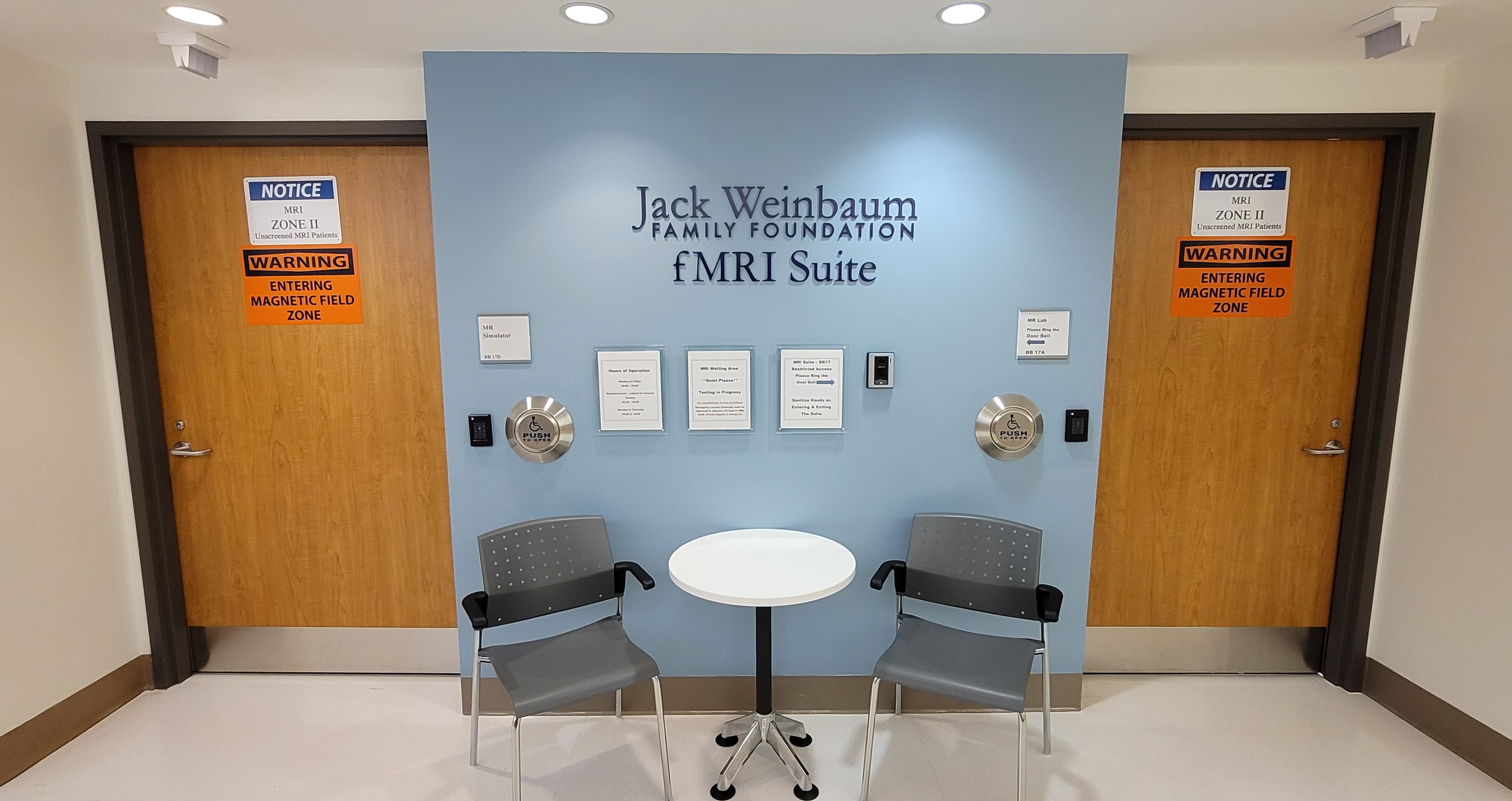
Renovations to the MRI Facility were completed in early 2021. The updated suite includes:
- Interview Room for participant screening
- Accessible Bathroom with Shower
- Change Room with lockers for personal possessions
- Simulator room with direct user-accessible entry from hospital hallway
- Control Room with plug-and-play stimulus equipment setup
- Large Scanner Room with cozy wood paneling
MRI Scanner
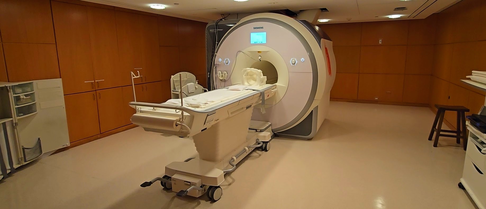
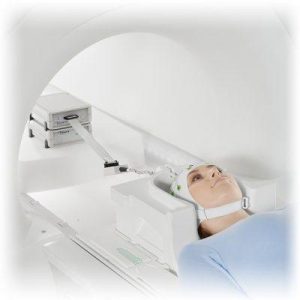
Our new Siemens 3T Prismafit dedicated research MRI Scanner was installed December 2020. The Prisma is Siemens’ latest research-focussed scanner, boasting an improved gradient strength of 80 mT/m and slew rate of 200 T/m/s, ideal for fast functional and diffusion imaging. We have three head coils available, a 20-channel head/neck coil, a 32-channel head coil, and a 64-channel head/neck coil, each suited to different studies and participants based on equipment selection and research goals. The scanner console software is currently XA30, with a full suite of product sequences from Siemens including recent developments like simultaneous-multi-slice (sms) EPI for functional and diffusion imaging, and 3D pseudo-continuous arterial spin labeling (3D pCASL) for perfusion imaging. We also have a variety of in-development and third party sequences available including multi-echo EPI for functional imaging of high susceptibility brain regions, and dual-echo arterial spin labeling (DE-ASL) for simultaneous perfusion and functional imaging.
Ancillary Equipment Overview
Stimulus Presentation

Computer
Dell Precision 5820
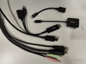
The Rotman MRI provides its users with a fully configured Stimulus PC. This PC is already wired to most of our equipment, so that no hardware setup is needed before each session. We have many popular stimulus softwares (including PsychoPy (python), E-Prime, Presentation, PsychToolBox (matlab)), as well as software tools specific to our other hardware systems (VPixx, SR Research), already installed on this computer. We maintain consistent software versions on this PC and install new versions in parallel to maintain consistent software versions for every project. Other software can be configured upon request. A bundle of KVM and hardware connected cables are available for researchers who wish to connect their own computers to our stimulus setup.
Visual Presentation System
Projector
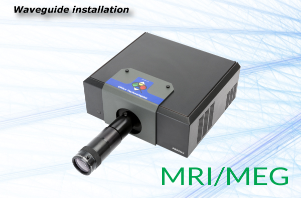
VPixx PROPixx MRI
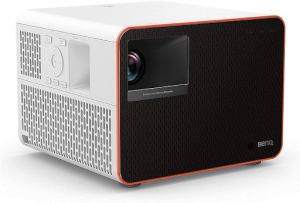
A full visual system from VPixx is newly installed at the Rotman, allowing high resolution (1920×1080), fast refresh rate (120Hz) visual stimuli, and the ability to precision synchronize other stimuli to the visual refresh rate for microsecond precision. Both our stimulus PC and KVM bundle include two separate video channels so that the stimulus display can be extended (default) or mirrored onto the projector display. This allows for separate researcher and participant displays, which can accommodate real time data validation, or stimulus scripts which maintain a consistent visual environment for the participant while stimulus software control is done only on the researcher display.
Audio Presentation System
Headphones
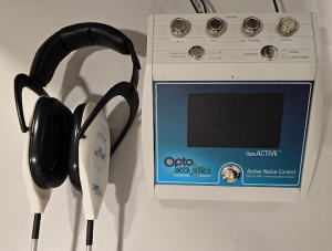
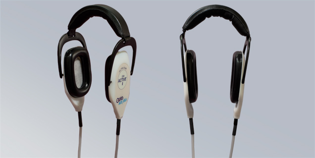
OptoAcoustics OptoActive Active Noise Cancelling Headphones
The OptoAcoustics system is our primary audio system, delivering exceptional audio clarity, compatible with all of our head coils, and capable of active noise cancellation of EPI scanner acoustic noise. Siemens loud-speaker audio presentation and communication are available for hardware combinations that are incompatible with over-ear headphones. All of our participants must use ear plugs regardless of audio system and project details. This provides a baseline passive audio decibel reduction that we deem necessary to protect our participants from loud scanner background noise.
Website: https://www.optoacoustics.com/medical/optoactive/features
Response System
Response Interface and Response Devices
Current Designs Birch Interface and assorted Response Devices
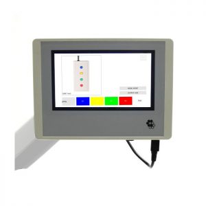
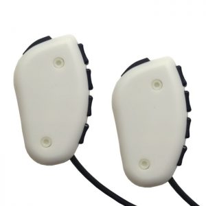
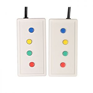
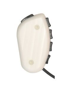
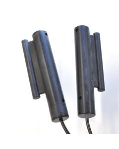
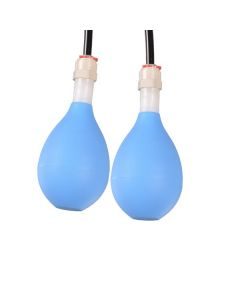
An updated participant response system from Current Designs includes a touch screen Birch interface and several response box options. This includes new ergonomic button boxes in addition to the legacy flat style boxes. User responses and scanner triggers are delivered as keyboard presses. Available response devices:
- Pyka – 8 Button Bimanual – Thumbs Disabled
- Legacy – 8 Button Bimanual – Curved Lines
- Pyka – 5 button Single Box
- Pneumatic Squeeze Ball Bimanual
- Grip Force Bimanual
Website: https://www.curdes.com/
Simultaneous Eye-Tracking
Eye-Tracker
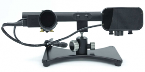
SR Research EyeLink 1000 Plus
A brand new SR Research EyeLink 1000 Plus was installed with the new scanner. We have mounted the camera on an external stand to prevent scanner vibrations from shaking the camera and affecting pupil tracking. We have also switched from battery packs to a permanent power solution. This means the eye-tracker is permanently set up, eliminating the need for camera placement and adjustments every session. Simply boot up the dedicated Host computer in the control room, which is already wired to the provided Stimulus Computer (SR software is already installed), adjust your thresholds, and you’re good to go.
Website: https://www.sr-research.com/solutions/fmri-meg-solutions/
Simultaneous fMRI-EEG
EEG
EGI MR-compatible Geodesic EEG System 400 OR Brain Products BrainAmp-MR EEG System
Two systems are available at the Rotman for simultaneous fMRI-EEG recording. The new EGI system uses 256-electrode saline-sponge caps which require ~15 minutes of setup and are suitable for most scanning sessions up to 1.5-2 hours. The Brain Products system uses 64 electrode gel caps which require 45-60 minutes of setup and are suitable for any length of scanning, including scanning above 1.5-2 hours such as MR sleep studies. Both systems are set up on mobile carts. This allows for the participant to be set up in another room (our simulator space is able to be reserved for this purpose) and moved into the MRI space at the time of scanner reservation. The long range optical and signal wiring are permanently installed in the MRI. Setup requires the researcher to place some hardware in/near the scanner, and make several cable connections in both the control room and the scanner room.
Website: https://www.egi.com/research-division/eeg-systems/mr-compatible-eeg-systems
https://www.brainproducts.com/solutions/application-fields/eeg-fmri/
Simultaneous Physiology Recording
Physiological Recording
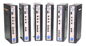
Biopac MP160 OR Siemens Built-In
A new Biopac MP160 and a variety of MRI Compatible modules enable recording of high quality physiological data during fMRI scans. Modules are available to measure Respiration, Pulse Ox, ECG, Skin Conductance, and O2/CO2 concentrations. Siemens also has some built-in transducers for recording Respiration, Pulse Ox, and ECG, although these produce lower quality data.
Website: https://www.biopac.com/application/magnetic-resonance-imaging-with-biopac-equipment/
MR Safe Corrective Lenses
Corrective Lenses
MediGlasses OR MRIFocus
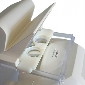
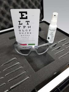
We have two sets of corrective lenses available at the Rotman MRI. The Mediglasses use interchangeable lenses that snap into an MR compatible frame that is worn by the subject within the head coil. The MRIFocus kit includes head coil specific lens mounts that interchangeable circular lenses sit in. It’s also possible to combine these two options for particularly strong prescriptions. An eye chart is available on site, set up at a viewing distance matching our scanner, for testing subjects who don’t know their prescription.
Website: https://wardray-premise.com/products/product/mr-mediglasses/
https://www.crsltd.com/tools-for-functional-imaging/vision-correction/mrifocus/
Mock (0T) MRI Simulator and Equipment
Psychology Software Tools Vera MRI Simulator and Stimulus Equipment
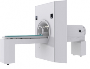
Our MRI Simulator is set up to match our real MRI environment on as many points as possible. The simulator has a functional patient table, a mock headcoil, and loud speakers for playing recorded MRI scanner noise. There is a matching stimulus computer or a set of KVM connections for using your own laptop. We have a projector with the same 1080p 120Hz specs as the VPixx. OptoAcoustics built us a custom non-MR headphone system with push-to-talk for realistic participant communication. For more details on the stimulus equipment, check our detailed equipment list at the top of this page.
